
Resources
The Neuropathology/Histochemistry Core is located in the Center for Neurodegenerative Disease (CND) on the 5th floor of the Whitehead Research Building. Approximately 1,000 sq. ft. of wet laboratory space is shared between the Neuropathology/Histochemistry Core laboratory and the CND brain bank facility.
The Neuropathology/Histochemistry Core laboratory is fully supplied with equipment needed for automated tissue processing, sectioning, histology, and immunohistochemistry. In addition, a dedicated brain bank room is equipped with five –80°C freezers for storage of frozen brain tissues. Two large refrigerators and several shelving units in a nearby cold room are available for storage of cryo-preserved tissues. A locked room on another floor contains shelving units designated for storage of formalin-fixed tissues.
Equipment
Tissue Processing and Embedding
Sakura Tissue-Tek VIP Tissue Processor
The Sakura Tissue-Tek VIP is an enclosed tissue processor which combines versatile programming with safe and efficient tissue processing. Processing cycle programs can be as diverse as required to meet the needs of the laboratory and the tissues being processed, and may range from short processing cycles of a few hours to extended or overnight programs.
Sakura Tissue-Tek TEC V Tissue Embedding Console System
The Tissue-Tek TEC V Tissue Embedding Console is an integrated system with all the components necessary for effective histological paraffin embedding. A temperature-controlled tank holds and dispenses molten paraffin as needed, an illuminated and temperature-controlled hot plate and small cooling plate are available for use as an embedding area, and a cold plate allows for cooling of embedded specimens prior to microtomy. 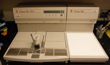
Additional heated storage spaces allow for heating and storage of embedding molds and tissue specimens prior to embedding.
Sectioning
Leica RM2235 Rotary Microtome
The Leica RM2235 Microtome is a precision engineered rotary microtome designed for general laboratory use in cutting paraffin sections. It is fitted with a cooling clamp to allow for constant cooling of the block being cut, and the precise clamp settings permit resumption of cutting of a previous block with minimum tissue loss. Moreover, the section thicknesses achievable range from 4 to 60 microns, providing the user with great versatility. 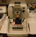
Shandon 0325 Microtome
The Shandon 0325 Microtome is a precision engineered rotary microtome designed for general laboratory use in cutting paraffin sections. Its unique “Accu-Feed” system allows for precision advancement of the specimen without the need for adjustment, resulting in more uniform paraffin sections. 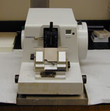
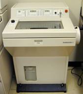 Shandon Cryotome SME
The Shandon Cryotome SME is designed for cutting frozen sections rapidly and accurately. It operates by rapidly freezing the specimen in a temperature-controlled environment, and allows for sectioning the frozen sample by conventional microtome. The SME combines automated, electronic specimen advance with independent temperature control of the specimen on the specimen head; the main body of the microtome is mounted outside the refrigerated chamber containing the specimen head to allow more working space.
Staining
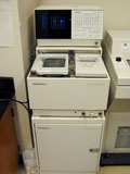
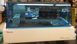
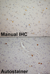
Thermo Scientific Autostainer 480S
The Autostainer 480S allows automation of nearly any manual immunohistochemistry procedure, while improving consistency and reproducibility via precise automation control. The system allows use of reagents from any source, and user-driven barcoding reduces errors. The antigen retrieval device uses gentle heat to produce outstanding, consistent results while maintaining the integrity of delicate tissues without the damaging effects of temperature variations, microwaves, or pressure.
Microscopy
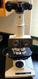
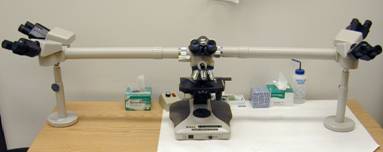
An Olympus BH-2 microscope is available for use in evaluation of specimens. The Neuropathology/Histo-chemistry Core also has access to a Nikon multi-headed microscope for shared viewing and discussion of results.
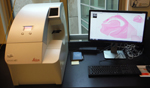
Leica Aperio AT2 Slide Scanner
The Aperio AT2 slide scanner is designed for speed and reliability in whole slide imaging. With its compact size, high capacity autoloader, and optimal image quality, it offers consistent, precise high-resolution capture of slide details for bright-field specimens. Images are available for remote viewing, allowing internal as well as external sharing and collaborations. There is also easy integration into laboratory information systems for image analysis and other applications.
Biobanking
Five -80°C freezers are currently in use for storage of frozen brain tissue samples. These are all on emergency generator back-up power and connected to an alarm system to notify staff in the event of a power failure or other emergency.
For more information on the resources utilized within this lab, please feel free to contact us